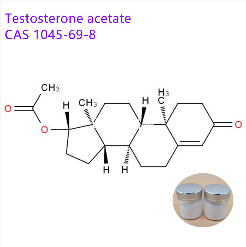
Steroid hormones, also known as steroid hormones, are a class of tetracyclic aliphatic hydrocarbon compounds with a cyclopentane polyhydrophenanthrene nucleus. It has very important medical value. It has a clear role in maintaining life, regulating sexual function, body development, immune regulation, skin disease treatment and birth control.
Steroids include steroids (eg cholesterol, lanosterol, sitosterol, stigmasterol, ergosterol), bile acids and bile alcohols, steroid hormones (eg adrenal corticosteroids, androgens, estrogens),
Our company specializes in providing steroid series products, welcome to inquire and order
Steroids Oil,Steroid Powder,Steroids Injections,Steroid Powder And Oil XI AN RHINE BIOLOGICAL TECHNOLOGY CO.,LTD , https://www.rhinebiotech.com
Since the concentration of the polyacrylamide gel can be formulated as required, two electrophoresis systems of "continuous system" and "discontinuous system" can be formed. The "discontinuous system" is characterized by a greatly improved resolution of sample separation. The main features of this electrophoresis are: (1) using two different concentrations of the gel system; (2) preparing the buffer composition and pH of the two gels, and the composition and pH of the electrophoresis buffer in the electrophoresis tank Not the same. In the experiment, the electrophoresis gel is divided into two layers: the upper layer of glue is a low concentration of macroporous glue, which is called concentrated glue or layered glue. The buffer for preparing the layer is Tris-HCl, pH 6.7; the lower layer of glue is It is a high-concentration small pore gel called separation gel or electrophoresis gel. The gelation buffer is Tris-HCl, pH 8.9. The electrode buffer in the electrophoresis tank is Tris-glycine, pH 8.3. It can be seen that the gel concentration, gelation composition, pH and electrophoresis buffer system are different, forming a discontinuous system.
During electrophoresis, the protein sample is placed on the concentrated gel. To prevent the protein sample from diffusing in the electrode buffer, an equal volume of 40% sucrose or 50% glycerol is added to increase the density; in order to observe the migration of the protein sample Tracer dyes such as bromophenol blue are also added to the sample. The molecules of these colored substances migrate faster than any macromolecular substance. As long as the dye does not move out of the gel tube, the sample does not run out of the hose.
In a discontinuous system, when the power is turned on to start electrophoresis, the glycine, protein, HCl, and bromophenol blue in the system are all dissociated into anions, forming an ion flow toward the anode. Its mobility depends on the number of charges, molecular weight and shape of the ions. However, when the glycine ions in the electrode buffer (pH 8.3) entered the concentrated gel, they encountered a lower pH (6.7) than pH 8.3, and the pH dropped by nearly two units, almost equal to the isoelectricity of glycine. Point (5.97), the degree of dissociation of glycine suddenly decreased, the amount of charge was significantly reduced, and the mobility was slowed down. The protein component in the serum sample also enters the concentrated gel. Although the pH change has an effect on the dissociation degree, it is much smaller than the glycine, the migration rate is larger than that of glycine, and the gel pore of the concentrated gel is larger for the protein. Molecules do not cause obstacles. The Cl- in Tris-HCl in the concentrated gel is completely dissociated, the molecular weight is small, the friction is not large, and the mobility is faster than that of protein and bromophenol blue. Thus, the mobility of various ions in the concentrated gel is formed: glycine < protein < bromophenol blue
When the glycine molecule enters the concentrated gel, the dissociation degree decreases, causing a sudden loss of the moving ion current, and the current decreases to decrease the conductivity. However, the current in other parts of the electrophoresis system remains unchanged, and is inversely proportional to the conductance and potential gradient (E=I/n, E is the potential gradient, I is the current intensity, and n is the conductivity), so the leader A high local potential gradient is suddenly formed between the ion Cl- ion and the slow ion glycine ion. The components of the serum protein in this local high-potential gradient region rapidly move forward to the Cl- ion region at different speeds (different molecular weights and different charge amounts) under the action of a high electric field. When the leading Cl- region is reached, the large electric field strength is weakened and the ion moving speed is rapidly slowed down. As a result, the protein sample between glycine and Cl- ions is stacked or concentrated into layers according to the size of the molecule. . Through this process, the protein sample is concentrated several hundred times, and the protein components are also arranged in layers in a certain order.
When the ion current continues to move forward into the small pore gel prepared in pH 8.9 buffer, the protein molecules encounter resistance in the small pore gel, the mobility is slowed down, and at the same time, the glycine is fully dissociated at pH 8.9. Its charge increases, eliminating the phenomenon of ion flux loss, and small molecules of glycine ions catch up with proteins. Each part of the gel recovers with a constant electric field strength, and the separation of the protein is carried out in a manner generally in the form of a general zone.
It can be seen from the above principle that the main advantage of the discontinuous electrophoresis of polyacrylamide gel is that after the protein sample is concentrated, a tightly compressed layer is formed into the separation gel. The components of the protein are separated and compressed into layers in advance, which can reduce the interference caused by the overlapping of the components due to free diffusion during electrophoresis, thereby improving the resolving power of electrophoresis. Due to this advantage, a small amount of protein sample (1-100??g) can also be separated well, and the high resolution enables the serum protein to obtain nearly 20 zones.
(2) Reagents and equipment
1. Sample serum proteins, desalted IgG, and purified IgG were tested.
2. Reagent
(1) Preparation of separation gel, concentrated gel related reagents
1 Gel buffer: Weigh 48 ml of 1 mol/L HCl, 36.6 g of Tris (trishydroxyaminomethane), 0.23 ml of TEMED (N, N, N`, N`-tetramethylethylenediamine), and add distilled water to 80 ml was dissolved, adjusted to pH 8.9, then the distilled water was added to a volume of 100 ml, placed in a brown bottle, and stored at 4 ° C.
2 Separation gel stock solution: 28% Arc-0.735% Bis stock solution: acrylamide (Acr) 28.0 g, methylidene bisacrylamide (Bis) 0.735 g, add distilled water to dissolve and dilute to 100 ml. After filtration, the brown reagent bottle is stored at 4 ° C, and can be placed for about 1 month.
3 Analysis of pure ammonium persulfate (AP) (AR) 0.14g of distilled water to 100ml, placed in a brown bottle, stored at 4 ° C can only be used for one week, the same day. The above three reagents are used to prepare a separation gel.
4 Concentrated gel buffer: Weigh 1ml/L HCl 48ml, Tris5.98g, TEMED 0.46ml, add distilled water to 80ml, adjust pH6.7, dilute to 100ml with distilled water, place in brown bottle, store at 4°C .
5 concentrated gel stock solution: called Acr10g, Bis2.5g, add distilled water to dissolve, dilute to 100ml, filtered and placed in a brown bottle, stored at 4 ° C.
6 40% sucrose solution (W/V)
7 riboflavin solution riboflavin 4.0mg, dissolved in distilled water, dilute to 100ml, placed in a brown reagent bottle, stored at 4 ° C.
(2) Ph8.3 Tris-glycine electrode buffer
Tris6.0g, glycine 28.8g, add distilled water to 900ml, adjust pH 8.3, dilute to 1000ml with distilled water, set in a reagent bottle, store at 4 ° C, diluted 10 times before use.
(3) 0.1% bromophenol blue indicator
(4) There are many types of dyeing liquids, and the dyeing methods are not identical. This experiment uses 0.05% Coomassie Brilliant Blue R250 staining solution, containing 20% ​​sulfosalicylic acid, which has the advantages of dyeing and fixing at the same time, and the background is easy to decolorize. 0.5% Coomassie Brilliant Blue R250 20% sulfosalicylic acid staining solution: Coomassie brilliant blue 0.05g, sulfosalicylic acid 20g, add distilled water to 100ml, and store in a reagent bottle after filtration.
(5) Decolorization solution 7% acetic acid solution
(6) 10 ml of glycerol in preservation solution, 7 ml of glacial acetic acid, and distilled water to 100 ml.
(7) 1% agar (sugar) solution 1 g of agar (sugar), add 10 times diluted electrode buffer, heat to dissolve, store at 4 ° C, set aside.
3. Equipment Electrophoresis instrument sandwich vertical plate electrophoresis tank.
(3) Operation method
1. Installing a sandwich vertical plate electrophoresis tank
The sandwich vertical plate electrophoresis tank is easy to operate and is not easy to leak. The two sides of the electrophoresis tank are electrode slots made of plexiglass, and a gel mold is sandwiched between the two electrode slots. The mold consists of an upper frame gel frame, long and short glass plates and a sample slot template (comb). Composed of. The electrophoresis tank consists of an upper storage tank (the platinum electrode is on or facing the short glass plate), a lower storage tank (the platinum electrode is below or facing the long glass plate) and a reticular condensation tube. The two electrode slots are fixed to the gel mold by a reservoir screw. The departments are assembled in the following order:
(1) Pin the upper storage tank and the fixing screws and place them on the table.
(2) Insert the upper and lower glass plates into the concave grooves of the upper frame-shaped silicone rubber. Be careful not to touch the glass of the glue surface by hand.
(3) Place the gel mold that has been inserted into the glass plate on the upper storage tank, and the short glass plate should face the upper storage tank.
(4) Align the pin hole of the lower tank with the upper tank where the screw pin is installed, and tighten the screw cap diagonally with both hands.
(5) Vertical electrophoresis tank, in which the melted 1% agar (sugar) is added in the gap between the lower end of the long glass plate and the silica gel mold frame. The purpose is to seal the voids, and bubbles should be avoided in the agar (sugar) after solidification.
2. Glue
1 Separation gel 20ml pH8.9 7.0% PAA solution, gel buffer (pH8.9) 2.5ml, separation gel stock 5.0ml, double distilled water 2.5ml, mix well, pump for 10min, then add AP 10 ml .
2 Concentrated gel pH 6.7 25% PAA: concentrated gel buffer: concentrated gel stock solution: 40% sucrose solution: riboflavin solution was mixed at a ratio of 1:2:4:1.
3. Preparation of gel plate
The discontinuous system uses separate gels and concentrated gels with different pore sizes and pH. The gel preparation should be carried out in 2 steps.
1 Preparation of the separation gel: According to the experimental requirements, the concentration of the final acrylamide is selected. In this experiment, 20 ml of pH 8.9 7.0% PAA solution is required, and the addition method is different from the continuous system. The mixed gel solution was added to the narrow slit between the long and short glass plates with a dropper of a slender head, and the glue height was about 1 cm from the lower edge of the sample template comb. A 1 cm syringe was used to gently add a layer of distilled water (about 3-4 mm) along the edge of the short glass plate to isolate the air to make the rubber surface flat. To prevent leakage, add distilled water slightly below the rubber surface in the upper and lower storage tanks. When the gel is completely polymerized for about 30-60 minutes, it can be seen that the water has a different refractive index from the solidified rubber surface. Use a filter paper strip to remove excess water, but do not break the rubber surface. If pre-electrophoresis is required, the distilled water in the upper and lower storage tanks is poured out, replaced with a separation gel buffer solution, and electrophoresed at 10 mA for 1 hour. After the electrophoresis is terminated, the separation gel buffer solution is discarded, and the number of the rubber surface is washed with a syringe. Once, the concentrated gel can be prepared.
2 Preparation of concentrated gel: The concentrated gel is pH 6.7 25% PAA. After mixing evenly, add the gel solution to the narrow slit of the long and short glass plates (that is, above the separation rubber) with a slender dropper, and the short glass plate At the upper edge of 0.5 cm, gently add the sample slot template. Distilled water is added to the upper and lower storage tanks, but cannot exceed the short glass upper edge. Photopolymerization was carried out at a distance of 10 cm from the electrode bath with a fluorescent lamp or the sun, but did not cause a large temperature. Under normal conditions, after 6-7 min of irradiation, the gel turned from egg yolk to milky white, indicating that polymerization started. Continue to illuminate for 30 min to complete the gel polymerization. After the light is collected and placed for 30-60 minutes, the sample cell template is gently taken out, and the excess liquid in the sample groove is sucked up with a narrow strip of filter paper, and a Tris-glycine electrode buffer diluted 10 times pH 8.3 is added. The liquid surface can be loaded without passing the glass plate by about 0.5 cm.
4. Loading
As a PAGE sample for analysis, only a few micrograms are needed, and after 2-3 ul of serum electrophoresis, dozens of protein bands can be separated. To prevent sample diffusion, an equal volume of 40% sucrose (containing a small amount of bromophenol blue) should be added to the sample. 5 ul of the above mixture was taken with a micro-syringe, and the sample was carefully added to the bottom of the gel-shaped sample tank through a buffer. After all the concave sample tanks were filled with the sample, electrophoresis was started.
5. Electrophoresis
Connect the positive pole of the DC stabilized electrophoresis instrument to the lower slot, and connect the negative pole to the upper slot (do not connect the wrong direction). Turn on the cooling water, turn on the electrophoresis switch, and initially adjust the current to 10 mA. The current was adjusted to 20-30 mA while the sample entered the separation gel. When the blue dye migrates to 1 cm from the lower edge of the rubber frame, the current is turned back to zero, and the power and cooling water are turned off. The upper and lower storage tank electrode buffers are separately collected in the reagent bottle, and stored at 4 ° C for 1-2 times. Loosen the fixing screw, take out the silicone rubber frame, gently remove a piece of glass plate with a stainless steel shovel, remove a corner at the end of the rubber plate as a mark, and move the rubber plate to a large petri dish for dyeing.
6. Fixed, dyed
In this experiment, 0.05% Coomassie Brilliant Blue R250 (containing 20% ​​sulfosalicylic acid) staining solution was used, and the staining and fixation were carried out simultaneously, so that the staining solution was not passed through the rubber plate and stained for about 30 minutes.
7. Decolorization
Rinse with 7% acetic acid for several times until the background blue faded. If you use a 50 ° C water bath or a faded shaker, the fading time can be shortened. After the decolorizing solution is decolorized by activated carbon, it can be used repeatedly. 


Protein Technology Topics: Polyacrylamide Gel Electrophoresis for IgG Purity Identification
(1) Principle